



Reconstructing the history of skeletal remains exposed to high temperatures is one of the most complex challenges in taphonomy, forensic science, and bioarchaeology. Heat induces profound, multiscale changes that alter the chemical composition, microcrystalline structure, and overall morphology of bone tissue.
A new exploratory study co-authored by Dr. Giulia Festa (researcher at the Enrico Fermi Research Center – CREF) alongside an international team of collaborators opens an innovative perspective in this field. The study, titled “New dimensions in burnt bone taphonomy through X-ray, neutron imaging and multivariate analysis”, was recently published in Scientific Reports (Vol. 16, Art. 33965).
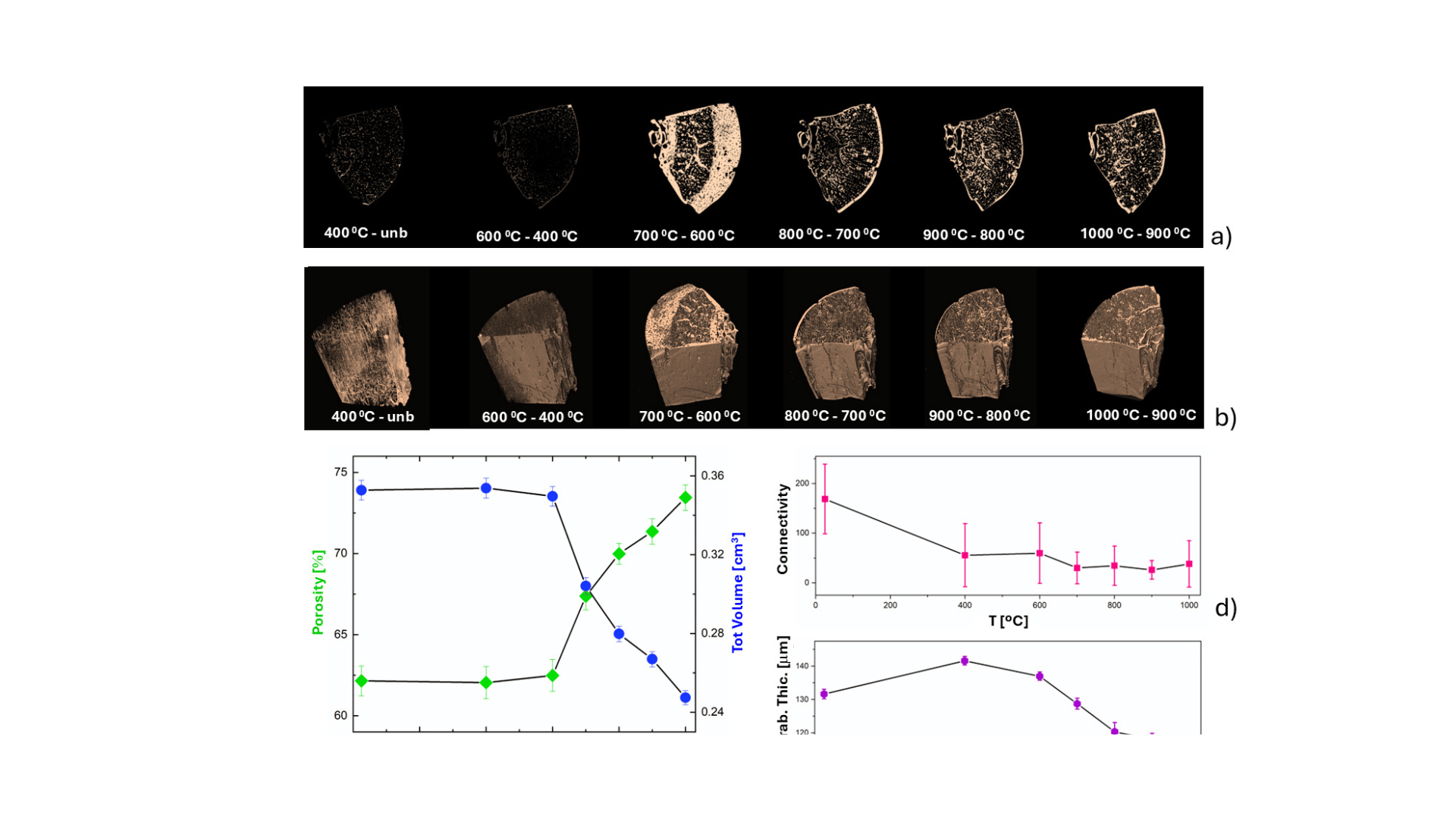
The scientific team used an integrated approach to analyze human femur samples (from a single donor) subjected to controlled heating in air across a temperature range from 25 °C to 1000 °C.
“The methodological innovation of this approach” said Giulia Festa “lies in the synergistic integration of two complementary, non-destructive imaging techniques. X-ray Tomography is ideal for evaluating density, three-dimensional geometry, while Neutron Tomography is particularly sensitive to light elements such as hydrogen, making it essential for tracking the loss of organic content and structural water within the bone as temperature increases.”
The datasets obtained from these imaging techniques were subsequently processed using multivariate statistical analysis, allowing the team to cross-reference chemical-crystallographic alterations directly with volumetric and micromorphological parameters. Using this framework, the researchers extracted and quantified several key structural parameters with high precision, demonstrating how thermal action progressively alters bone architecture, creating a specific “fingerprint” for each temperature regime.
“This research is a powerful tool for Bioarchaeology and Anthropology, but also for Forensic Sciences” said Dr Festa. “delivering objective evidence for investigations, such as estimating the maximum temperature reached in different scenarios.
This publication reinforces CREF’s role as a leading research hub in applying advanced physical methodologies—specifically neutron techniques and advanced imaging—to solve complex problems across cultural heritage, anthropology, and forensic medicine.
Article Details
- Title: New dimensions in burnt bone taphonomy through X-ray, neutron imaging and multivariate analysis
- Authors: G. Festa, M. P. M. Marques, L. A. E. Batista de Carvalho, S. Nonni, A. L. C. Brandão, D. Gonçalves, F. Leonard, L. Sinclair, D. Duggins, S. F. Parker & W. Kockelmann
- Journal: Scientific Reports (2026), Volume 16, Article number: 33965
- Link: Scientific Reports – Article 33965